New publication: MRI-based neuroanatomy of the Baird’s beaked whale brain

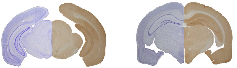
We are pleased to share our new publication on the brain anatomy of the Baird’s beaked whale (Berardius bairdii), one of the largest toothed whales and a rarely studied member of the beaked whale family, Ziphiidae.

Using magnetic resonance imaging (MRI), we provide a detailed description of the external and internal brain anatomy, including cortical folding, major brain structures and volumetric measurements. The Baird’s beaked whale brain shows a typical odontocete organization, with a large and highly folded neocortex.

At the same time, the encephalization quotient and relative cerebellar volume are lower than in delphinids, consistent with findings in other deep-diving cetaceans. These differences may reflect adaptations related to diving behavior, large body size and energetic constraints.

Join Us for the Summer School

Nina will teach a summer school at Hokkaido University, Japan, organized by Prof. Khin Khin Tha.

This interdisciplinary course integrates neurobiology and quantitative MRI (qMRI) to explore how brain structure and function can be objectively characterized. Through eight interactive sessions combining lectures, discussions, and student presentations, participants will connect imaging-derived biomarkers with underlying tissue structure and physiology.

The course emphasizes interdisciplinary learning, linking anatomy, physics, and clinical imaging to foster advanced understanding of brain organization and promote international academic collaboration.

Eligibility: Current students and non-students
Level: Graduate
Format: On-campus or online

Registration and further information: [Link]

Cortical neuronal numbers in the Northern minke whale align with artiodactyl scaling rules

In our newly published study we analyzed the neuronal composition of the Northern minke whale brain. We find that the cerebral cortex, despite being about twice the mass of the human cortex, contains only the number of neurons expected for an artiodactyl brain of its size, and far fewer than in humans and great apes. At the same time the minke whale shows an unusually high proportion of cerebellar neurons relative to cortical neurons, a specialization that may be linked to their unique acoustic communication. These results advances our understanding of cetacean brain composition and highlight conserved as well as unique aspects of their neural architecture. Read the full study here [Link].

BSAF Award for Best Neuroimaging Paper 2025

We are happy to share that our paper “Anatomical and volumetric description of the Guiana dolphin (Sotalia guianensis) brain from an ultra-high-field magnetic resonance imaging”  received the BSAF Best Neuroimaging Award.

The study provides the first comprehensive anatomical and volumetric description of the Guiana dolphin brain using ultra-high-field MRI. These data offer an important reference point for future comparative and evolutionary neuroanatomy research.

The article is freely available: [Link].

Read Our Commentary in the Journal of Comparative Anatomy: “Of Marine Mammal Neuroscience and Men”Needs and Perspectives in Marine Mammal Neuroscience

In our recent commentary, we highlight the urgent need to advance marine mammal neuroscience—a field that, despite the rapid evolution of neurotechnologies, remains underdeveloped and fragmented. Cetacean and pinniped brains show remarkable evolutionary specializations, but systematic, reproducible data across species remain scarce due to constraints in sampling, access, and methodological standardization.

We identify a critical need for coordinated efforts to standardize best practice protocols for the sampling, storage, and systematic analysis of marine mammal nervous tissues. To this end, we propose the formation of an inclusive, multidisciplinary network and invite collaboration through our Open Science Framework project.

Join us to collectively advance this emerging field!